

Scientific-Technical Services
Textural and Thermal Analysis Service
Textural and Thermal Analysis Service
Thermal Analysis
 |
Scientific responsible Dr. Luis A. Pérez Maqueda · Dr. Pedro Enrique Sánchez Jiménez Technical staff Dª Cristina Gallardo López contact ✉ maqueda@icmse.csic.es ☎ 954 48 95 48 |
Thermal analysis techniques allow to studying physical or chemical changes occurring in solid in samples as a function of the temperature. Those changes should involve either a mass change or a heat flow.
The experiments can be performed in the range from room temperature to 1500ºC, both under inert (N2), or reactive (air, O2,…) atmospheres.
Two different techniques are available: Thermogravimetry (TG) and Differential Thermal Analysis (DTA).
Available Equipment
- STA449 F5 Jupiter (NETZSCH) Simultaneous TG/DTA/DSC Instrument
- DIL 402 Expedis Select (NETZSCH) Horizontal Dilatometer
- TA Instruments Q600 Simultaneous TG/DTA/DSC instrument
- Thermogravimetric instrument TG, TA Instruments Q5000
- Calvet Calorimetry Equipment, Setaram Sensys
|
Services
|
External | |
|---|---|---|
| OPI / AGE / Universidades | Others | |
| Differential Thermal Analysis and Thermogravimetry DTA-TG | 200,00 (€/sample) | 219,04 (€/sample) |
Physisorption-Chemisorption
 |
Scientific responsible Dr. Gerardo Colón Ibáñez · Dr. Alfonso Caballero Martínez Technical staff Dª Cristina Gallardo López contact ✉ gcolon@icmse.csic.es · caballero@us.es ☎ 954 48 95 36 - 954 48 95 43 |
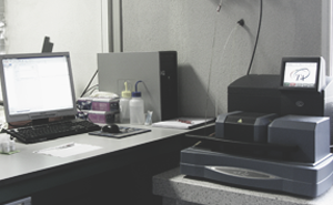
This service constitutes a basic tool for the microstructural characterization of powdered solids of different natures, regarding to their porosity, specific surface area and chemically active surface.
This service is composed by a phyisisorption analyser (Micromeritics, ASAP 2020) which pro-vides the complete adsorption/desorption isotherms, from which the specific surface area, pore and micropore size distribution and concentration of reactive sites are obtained. The instrument is also equipped for carrying out chemisorption of different reactive molecules, as O2, H2, CO, etc.
Available Equipment
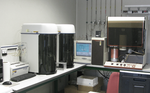
- Physicorption Analyser ASAP2010 (Micromeritics)
- Chemisorption Analyser ASAP2010 (Micromeritics)
- Multisample Physisorption Analyser TRISTAR II (Micromeritics)
- Multisample Physisorption Analyser TRISTAR II-Kr (Micromeritics)
|
Services
|
External | |
|---|---|---|
| OPI / AGE / Universidades | Others | |
| Temperature Programmed Desorption (TPD-CO2) | 89,30 (€/sample) | 97,80 (€/sample) |
| Temperature Programmed Oxidation (TPO-O2) | 89,30 (€/sample) | 97,80 (€/sample) |
| Chemisorption H2 | 89,30 (€/sample) | 97,80 (€/sample) |
| Temperature Programmed Reduction (TPRH2) | 89,30 (€/sample) | 97,80 (€/sample) |
| Specific Surface Area (BET) | 89,33 (€/sample) | 97,84 (€/sample) |
| Superficie específica (Isoterma Adsorción-Desorción) | 112,72 (€/sample) | 123,46 (€/sample) |
Atomic Emission Spectrometry
Atomic Emission Spectrometry
Atomic Emission Spectrometry (ICP-OES)
 |
Scientific responsible Dr. Francisco José Gotor Martínez Technical staff Lda. Belinda Sigüenza Carballo contact ✉ belinda@icmse.csic.es ☎ 954 48 95 39 · int.: 446139 |
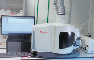
Inductively coupled plasma atomic emission spectrometry (ICP-OES) is an analytical technique that allows the quantification of elements up to the trace level in samples in solution. The sample to be analyzed is nebulized and conducted to an argon plasma, where desolvation, vaporization, atomization and ionization of the elements take place. The atoms and ions reach an excited state by the high thermal energy supplied by the plasma and during the relaxation process electromagnetic radiation is emitted with wavelengths characteristic of each element. The intensity of the different emission lines is proportional to the concentration of the elements, which can be quantified by using appropriate calibration curves. This technique has high detection limits (in the ppb range, μg / L), good reliability, high throughput and multi-elemental capacity, although in some cases spectral interferences can occur due to a high number of emission lines.
Solid samples (digestion will be carried out by the service) or liquid samples in slightly acidic aqueous solution can be supplied. Samples in HF medium are not allowed. Liquid samples must not present precipitates or colloids in suspension and must have a minimum volume of 10 ml. The samples will be delivered to the technician in charge of the service, together with the duly completed analysis request that is available on the ICMS website.
Available Equipment
- iCAP 7200 ICP-OES Duo (ThermoFisher Scientific)
- Microwave Digestion System ETHOS EASY (Milestone)
|
Services
|
External | |
|---|---|---|
| OPI / AGE / Universidades | Others | |
| Analysis of elements: ICP-OES. Análisis Elemental ICP-OES Muestras Líquidas | 22,27 (€/element) | 24,39 (€/element) |
| Digestión de Muestra | 32,11 (€/sample) | 35,17 (€/sample) |
Visible, ultraviolet, infrared and Raman Spectroscopy Service
Visible, ultraviolet, infrared and Raman Spectroscopy Service
Infrared Spectroscopy
 |
Scientific responsible Dr. Mauricio Calvo Roggiani · Dr. Miguel Angel Centeno Gallego Technical staff Dr. Miguel Angel Avilés Escaño contact ✉ mauricio.calvo@icmse.csic.es · centeno@icmse.csic.es ☎ 954 48 95 39 |
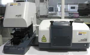
Infrared spectroscopy (FT-IR) is based on the selective absorption of the infrared radiation by the materials. This absorption means a change in the vibrational energy of the chemical bonds, whenever it occurs a change in the polarization. The result is a spectrum showing the absorbed or transmitted radiation as a function of the wavenumber of the radiation, which can be assigned to the corresponding chemical bound.
The equipment at the ICMS works in a wavenumber range from 5000 to 250 cm-1 (CsI optic), and can operate with a gas purge or in vacuum. It is equipped with several accessories to do Diffuse Reflectance (DRIFT), Attenuated Total Reflectance (ATR) or Specular Reflectance. It has got an Infrared Microscope with a lateral resolution of 10 µm.
Available Equipment
- JASCO FT/IR-6200 IRT-5000
|
Services
|
External | |
|---|---|---|
| OPI / AGE / Universidades | Others | |
|
Spectroscopy: FT-IR
|
52,25 (€/hour)
|
57,23 (€/hour)
|
Micro-Raman Spectroscopy
 |
Scientific responsible Dr. Miguel Angel Centeno Gallego · Dr. Mauricio Calvo Roggiani Technical staff Dr. Miguel Angel Avilés Escaño contact ✉ centeno@icmse.csic.es · mauricio.calvo@icmse.csic.es ☎ 954 48 95 43 - 954 48 95 81 |
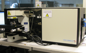
Raman spectroscopy is based on a photonic process in which the incident radiation is dis-persed by the sample. This latter is perturbed leading to vibrational and rotational transitions. In general, the Raman spectrum is interpreted like a vibrational one, providing information very similar to the infrared spectroscopy, although the Raman active vibrations are not always the same as those excited with infrared radiation. A Raman vibration mode is active if there is a change of polarizability of the chemical bonds or the considered molecule, which in turn results in the generation of induced dipolar momentam. Its application fields are very broad: semiconductors, carbon compounds (graphite, diamond, nanotubes, fibers…), catalysts, pigments, etc.
Available Equipment
- LabRAM Horiba Jobin Yvon equipped with a confocal microscope and 3 excitation lasers (785 nm red, 532 nm green, and 325 nm UV)
|
Services
|
External | |
|---|---|---|
| OPI / AGE / Universidades | Others | |
|
Micro-Raman Spectroscopy
|
52,60 (€/hour)
|
57,61 (€/hour)
|
Electroluminescence Spectroscopy
 |
Scientific responsible Dr. Mauricio Calvo Roggiani · Dr. Hernán Míguez García contact ✉ mauricio.calvo@icmse.csic.es · h.miguez@csic.es |
The ICMS electroluminescence spectroscopy service allows full characterization of current-activated photoemission from an emitting device. The service has an integrating sphere with a port that houses the electroluminescent device connected to a CCD detector. A module for angular characterization of the electroluminescent device is also available. The measurements allow to obtain the luminance vs current density curves or vs potential and the quantum efficiency of electroluminescence.
Available Equiment
- Measuring EQE System: C9920-12
- Light Brightness Distribution: C9920-11
|
Services
|
External | |
|---|---|---|
| OPI / AGE / Universidades | Others | |
| Spectrophotometry and Photoluminescence | 186,94 (€/hour) | 186,94 (€/hour) |
Ultraviolet-Visible-Near Infrared Spectroscopy
 |
Scientific responsible Dr. Gabriel Sebastián Lozano Barbero · Dr. Hernán Míguez García Technical staff Dr. Miguel Angel Avilés Escaño contact ✉ glozano@icmse.csic.es · h.miguez@csic.es ☎ 954 48 95 39 |
The Ultraviolet-Visible-Near Infrared Spectroscopy (UV-Vis-NIR) reports on the existing energy differences between the more external occupied electronic levels and the nearer unoccupied ones.
There are two equipments in the laboratory, which work in the wavelength range of 190 nm to 900 nm. It can operate in the Transmission mode or in Diffuse Reflectance Modes.
Available Equipment
- Cary 5000+UMA (Universal Measurement Accessory)
- Cary 300
|
Services
|
External | |
|---|---|---|
| OPI / AGE / Universidades | Others | |
|
Spectroscopy: UV-Visible Spectral Reflectometry (CARY 300)
|
49,28 (€/hour)
|
65,70 (€/hour)
|
| Spectroscopy: UV-Visible Spectral Reflectometry-NIR (CARY 7000) |
70,01 (€/hour)
|
106,68 (€/hour)
|
Ultrafast emission and absorption spectroscopy
 |
Scientific responsible Dr. Hernán Míguez García · Dr. Juan F. Galisteo López contact ✉ juan.galisteo@csic.es · h.miguez@csic.es ☎ 954 48 95 87 |
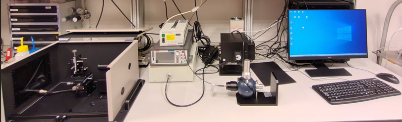
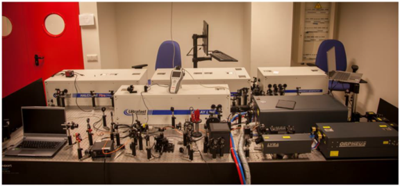
The ultra-fast spectroscopy laboratory allows performing time-resolved absorption and emission measurements with a time resolution of 190 femtoseconds (fs) over a broad temporal range (190 fs - 1 millisecond). Measurements can be carried out in the 350-850nm spectral range.
Instrumental disponible
- Sistema de excitación láser ultra-rápido formado por un láser pulsado PHAROS (Light Conversion) (longitud de onda de emisión 1030nm, tasa de repetición 1kHz y duración de pulso 190fs) y un amplificador paramétrico (OPA) ORPHEUS (Light Conversion) que produce pulsos de duración y tasa de repetición iguales al PHAROS pero con una longitud de onda sintonizable en el rango 350-2500nm.
- Espectrómetros de absorción para el rango temporal 190fs-8ns (HELIOS, Ultrafast Systems) y 2ns-1ms (EOS, Ultrafast Systems). Ambos sistemas permiten realizar medidas en el rango espectral 350-1100nm con una resolución de 2nm.
- Espectrómetro de emisión para el rango temporal 190fs-5ns (HALCYONE, Ultrafast Systems) oeprativo en el rango espectral 350-1100nm.
- Sistema de time-correlated single-photon counting (TCSPC) para realizar medidas de emisión resuelta en el tiempo en el rango temporal 1ns-1ms y en el rango espectral 200-850nm.
|
Services
|
External | |
|---|---|---|
| OPI / AGE / Universidades | Others | |
| Ultrafast Emission and Absorption Spectroscopy | 488,02 (€/h) | 488,02 (€/h) |
Surface Analysis Service
Surface Analysis Service
Chemical Surface Analysis by Electron Photoemision
 |
Scientific responsible Dr. Juan Pedro Espinós Manzorro · Dr. Juan Pedro Holgado Vázquez Technical staff Lda. Verónica Rodríguez Bravo · Gdo. Antonio González Moreno contact ✉ jpespinos@icmse.csic.es · holgado@icmse.csic.es ☎ 954 48 95 30 - 954 48 95 36 |
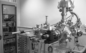
Typically, “photoelectron spectroscopies” are a powerful set of non-destructive analysis techniques, exclusively sensitive to the more superficial few atomic layers (20-30 Å), allowing to obtain valuable information about their chemical, physical and electronic properties.
The technical interest of the resulting information is huge in fields such as catalysis, corrosion, surface treatments,, floating and adhesion phenomena, or segregation processes, among others. The most remarkable characteristic of X-Ray Photoelectron Spectroscopy (XPS/ESCA) is that it allows to discriminate, for a given element, between different oxidation states or chemical surroundings (coordination).
Available Equipment
Photoelectron Spectrometer PHOIBOS 100-DLD, consisting on:
- Analysis Chamber, equipped with a hemispheric multichannel analyser PHOIBOS 100-DLD, a four axis manipulator, a dual X-ray source (achromatic Al Kα, Mg Kα), a UV lamp, and a electron gun, allowing to perform surface analysis by XPS, UPS, ISS and REELS, including angular resolved studies.
- Two prechambers for different treatments, with ultimate vacuum levels of 10-8 and 10-9 mbar respectively, where samples can be subjected to diverse treatments and transferred to the analysis chamber without exposure to the atmosphere. The possible treatments include heating at high temperature (< 800C) under controlled atmosphere, ion sputtering with inert or reactive gases, exposure to plasma, laser treatments, deposition of metals, oxides or simple compounds, exfoliation, etc.
Photoelectron Spectrometer SPECS, consisting on:
- Analysis Chamber, equipped with a hemispheric multichannel analyser PHOIBOS 100, three axis manipulator and dual X-ray source (achromatic Al Kα, Mg Kα).
- Pre-chamber for High Pressure/High Temperature treatments (HPHT Cell).Samples can be subjected to treatments in the presence of gases up to 20 bar and 800ºC (simultaneously). These treatments can be performed either under static or flowing gas conditions. After treatments, samples can be transferred to the analysis chamber without exposure to the atmosphere.
- A Fast entry chamber, equipped with a parking and degassing system, allowing the samples to be evacuated at moderate temperature (T < 150ºC). It is also possible to sputter the samples under an accelerated ion beam (0.5- 5.0 kV) using inert or reactive gases. Incorporation of some other equipment (Visible light illumination, metal evaporators) is also contemplated.
|
Services
|
External | |
|---|---|---|
| OPI / AGE / Universidades | Others | |
|
Spectroscopy Ultraviolet Photoelectron Spectroscopy (UPS). Análisi de UPS o REELS o ISS con Técnico
|
132,45 (€/hour)
|
145,06 (€/hour)
|
|
Spectroscopy X-ray Photoelectron Spectroscopy (XPS). Análisis XPS con Técnico
|
135,43 (€/hour)
|
148,33 (€/hour)
|
| Spectroscopy X-ray Photoelectron Spectroscopy (XPS). Tratamiento in situ con Técnico | 135,43 (€/hour) | 148,33 (€/hour) |
| Spectroscopy X-ray Photoelectron Spectroscopy (XPS). Análisis Cuantitativo de los Datos con Técnico | 179,65 (€/hour) | 196,75 (€/hour) |
| Spectroscopy X-ray Photoelectron Spectroscopy (XPS). Emisión de Informe Científico | 199,44 (€/hour) | 218,44 (€/hour) |
Contact Angle and Surface Tension Determination Service
 |
Scientific responsible Dra. Carmen López Santos contact ✉ mclopez@icmse.csic.es |
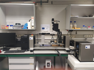
Advanced characterization of wetting using a goniometer to determine the contact angle of liquid droplets deposited on a surface is a quantitative and non-destructive analysis technique in general, depending on the surface reactivity with the wetting liquid. The combination of several liquids allows to determine the surface tension of the wet solid and to estimate the work of adhesion in simulated environments. The control of the wettability of smart surfaces under the action of external stimuli such as the application of light, temperature and/or humidity gradients, changes in pH or electric fields, are of great interest in advanced applications for the aeronautical industry, lines of communication, transportation, protection, heritage, solar and wind energy or biomedicine, among others.
Determination of the contact angle is carried out by applying Young's equation for interfacial tension balance, with sensitivity at the nanometric scale. The tests can be carried out in static or in dynamic modes to obtain hysteresis (advanced and receding angle values) of surfaces and critical values of droplet sliding. Regarding the surface tension of the solid surface, implemented theoretical models are those of Owens, Wendt, Rabel and Kaelble, Extended Fowkes, Schultz and Van Oss&Good and Neumann, based on the existence of polar and dispersive components. In addition, the temporal tracking of the surface contact angle accounts for aging and stability in certain environments. Finally, the assistance of a controlled temperature and humidity chamber allows studies of condensation/evaporation and freezing/thawing of water on surfaces.
The contact angle and surface tension measurement equipment has been supported by the Junta de Andalucía (Aid for R&D infrastructure and equipment 2019-PAIDI2020 European Regional Development Fund)
Available Equipment
DataPhysics OCA 25 Optical Contact Angle Goniometer, consisting of::
- double injection system with liquid drop volume, between nanoliters (close to the state of the art) to tens of microliters, controlled by a magnetic valve for dispensing
- TBU100 tilting platform for studies of droplet sliding on the surface (-5º - 95º)
- sample holder base adjustable in 3 spatial directions
- TPC160 thermoelectric module for temperature control (from -30ºC to 150ºC)
- module for the application of electric field EWP100 in studies of wetting electrostimulation (0-64kV DC/AC, 3-10mm electrode distance)
- HGC30 electro-humidification chamber to control the humidity of the environment (from 5% to 90%)
- SCA 21 software with routine for surface tension estimation (0.01-2000 mN/m)
- front (3250 f/s) and vertical (2450 f/s) optical systems
|
Services
|
External | |
|---|---|---|
| OPI / AGE / Universidades | Others | |
|
Medida del Ángulo de Contacto en Superficie
|
57,89 (€/hour)
|
63,41 (€/hour)
|
|
Medida del Ángulo de Contacto en Superficie (Electrowetting )
|
59,64 (€/hour)
|
66,32 (€/hour)
|
|
Medida del Ángulo de Contacto en Superficie (Wetting Ambiente Controlado)
|
110,35 (€/hour)
|
110,35 (€/hour)
|
Tribological and Mechanical Surface Characterization Service
 |
Scientific responsible Dr. Juan Carlos Sánchez López Technical staff Lda. Belinda Sigüenza Carballo contact ✉ jcslopez@icmse.csic.es |
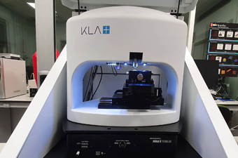
The KLA G200x nanoindenter enables the evaluation of the mechanical properties in the nanoscale (hardness, Young’s modulus) which results crucial for thin films and surface treatments, discarding the substrate influence. The CSM (Continuous Stiffness Measurement) option allows a continuous measurement of the stiffness as a function of the frequency or penetration. This operational mode results very useful not only for rigid materials as metals, ceramic or alloys, but also for materials with mechanical response dependent on time, like polymers, composites or biomedical.
The equipment disposes of two actuators (low and high load) that cover forces range up to 50 and 500 mN, respectively.
Additionally, the service includes the possibility of recording the 3D-surface properties by optical and interpherometric microscopy to evaluate the topography and roughness of the initial state of specimens and after being submitted to mechanical and tribological essays.
Available Equipment
- Nanoindenter KLA G200x
- 3D-optical profiler confocal and interferometric SENSOFAR S-Neox
|
Services
|
External | |
|---|---|---|
| OPI / AGE / Universidades | Others | |
|
Mechanical Properties of Materials at Microscale
|
209,16 (€/hour)
|
229,08 (€/hour)
|
|
3D-Optical Microscopy Confocal/High Resolution Perfilometry/Interferometry
|
124,72 (€/hour)
|
136,59 (€/hour)
|
X-Ray Diffraction Service
X-Ray Diffraction Service
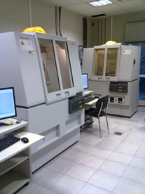
X-Ray Diffraction Laboratory
 |
Scientific responsible Dra . Ana Isabel Becerro Nieto (anieto@icmse.csic.es) Technical staff Gdo. Ángel Arias Pérez contact ✉ difraccion@icmse.csic.es ☎ 954 48 95 45 |
X-ray diffraction allows the qualitative and quantitative identification of crystalline substances and their microstructural and textural characterization.
At present, four independent diffractometers are available in this service, specifically config-ured to analyze the composition, chemical stability, crystallinity and many other properties in polycrystalline samples of a varied nature. Besides ordinary analyses (Θ-2Θ), part of the equipment can perform some advanced studies as:
- Direct monitoring of transformations undergone in materials under heating, such as phase changes, under inert o reactive atmosphere.
- To characterize materials at the nanoscale (1-100 nm) through X-ray scattering at low angles, using the SAXS technique.
- To measure some physical parameters of layers such as density, thickness and surface rough-ness with the reflectometry setup.
- To obtain the diffraction patterns of samples either sensitive to the atmosphere or highly transparent to X-rays (organic compounds) employing the capillary configuration.
Available Equipment
- Diffractometer PANALYTICAL X’PERT PRO with automatic sample charger
- Diffractometer PHILIPS X'PERT with high temperature chamber (1200ºC) ANTON PAAR HTK 1200
- Diffractometer PANALYTICAL X’PERT PRO (reflectometry, SAXS, low angle scattering and capillary)
|
Services
|
External | |
|---|---|---|
| OPI / AGE / Universidades | Others | |
| X-ray Scattering: Thin Film Characterization (Reflectivity, Reciprocal Space Maps) (Textura Empyrean) | 234,20 (€/hour) | 379,19 (€/hour) |
| Structural Characterization: Qualitative X-ray powder diffraction analysis (Cargador) | 22,70 (€/hour) | 36,76 (€/hour) |
| Structural Characterization: Qualitative X-ray powder diffraction analysis (Kalfa1) | 270,87 (€/hour) | 438,55 (€/hour) |
| Structural Characterization: Qualitative X-ray powder diffraction analysis (Microdifracción) | 269,93 (€/hour) | 437,03 (€/hour) |
| Structural Characterization: Qualitative X-ray powder diffraction analysis (X-Ray Diffraction with Temperature Chamber) | 18,58 (€/hour) | 30,09 (€/hour) |
| Small Angle X Ray Scattering (SAXS) | 24,72 (€/hour) | 40,03 (€/hour) |
| (X-Ray Diffraction) of thin films (Empyrean) | 147,68 (€/hour) | 239,11 (€/hour) |
Electron Microscopy Service
Electron Microscopy Service
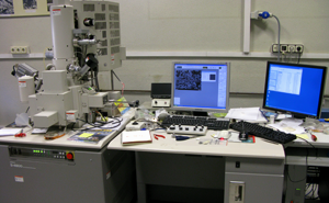
Scanning Electron Microscopy
 |
Scientific responsible Dra. Cristina T. Rojas Ruiz · Dr. Asunción Fernández Camach Technical staff Dra. Mª Carmen Jiménez de Haro contact ✉ tcrojas@icmse.csic.es · cjimenez@icmse.csic.es · asuncion@icmse.csic.es ☎ 954 48 96 25 |
The scanning electron microscopy provides information about the microstructure, morphology and chemical composition at the microscopic scale of solid samples. It can be applied to all type of materials including ceramics, polymers, metals, minerals, catalysts, samples from cultural heritage, thin films, coatings, interfaces, nanoparticles, etc. The SEM microscope is a field emission cold cathode equipment which enables images of the surface morphology and texture of samples with a resolution of 1 nm at 15kV. It also allows working at low voltages with non-metalized samples and in transmission mode for electron-transparent samples (STEM-in-SEM). Coupled to the X-ray detector (EDX) enables compositional analysis and elemental mapping.
Available Equipment
- Hitachi S4800 SEM-FEG microscope: cold cathode field emission gun with voltage from 0.5 to 30 kV, resolution of 1nm at 15 kV. Equipped with a Bruker-X Flash-4010 EDX detector with a resolution of 133 eV (at the MnKα line), and a detector with sample holder to work in transmission mode (STEM-in- SEM).
- Additional equipment in the “electron microscopy samples preparation laboratory”
|
Services
|
External | |
|---|---|---|
| OPI / AGE / Universidades | Others | |
|
Metal Coater (Sputtering)
|
41,20 (€/10 samples)
|
45,12 (€/10 samples)
|
|
Scanning Electron Microscopy (SEM): High Resolution SEM
|
71,18 (€/hour)
|
88,13 (€/hour)
|
|
Scanning Electron Microscopy (SEM): Low Voltage SEM
|
71,18 (€/hour)
|
88,13 (€/hour)
|
|
Scanning Electron Microscopy (SEM): EDS(Energy Dispersive X-Rays Spectroscopy)
|
71,18 (€/hour)
|
88,13 (€/hour)
|
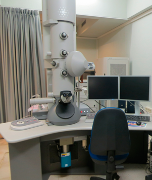
Transmission Electron Microscopy
 |
Scientific responsible Dra. Cristina T. Rojas Ruiz · Dra. Asunción Fernández Camacho · Dr. Miguel Anaya Martín Technical staff Dra. Luisa María Valencia Liñán · Lda. Olga Montes Amorín · Dª. Inmaculada Rosa Cejudo contact ✉ tcrojas@icmse.csic.es · olga@ciccartuja.es · asuncion@icmse.csic.es ☎ 954 48 96 25 |
The transmission electron microscopy is a widely used technique for the microstructural and chemical characterization at micro and nanoscales, providing two-dimensional images of the sample texture and shape as well as grain and/or particle size, degree of homogeneity at the microscopic scale, degree of crystallinity of the sample, identification of crystalline phases, and high resolution images to identify the crystalline domains. The microscope is equipped with an EDX analyzer for compositional analysis. It can be applied to all type of materials and research topics in materials science and technology working with electron-transparent samples prepared ad-hoc for this end. The service performs transmission electron microscopy: Imaging in bright and dark field, selected area electron diffraction and high resolution electron microscopy, as well as elemental analysis of selected areas. It does not provide STEM mode.
Available Equipment
- JEOL 2100Plus microscope (200kV) with LaB6 filament. Structural resolution of 0.14 nm between lines and 0.23 nm between points. Sample holders with one and two angles. An X-ray Energy Dispersive Analyzer (EDX X-Max 80T, Oxford Instruments) and a CCD camera (Gatan) for image registration are attached to the equipment.
- Additional equipment in the “electron microscopy samples preparation laboratory”
|
Services
|
External | |
|---|---|---|
| OPI / AGE / Universidades | Others | |
|
Structural Characterization: Tranmision Electron Microscopy (TEM)
|
66,75 (€/hour)
|
82,64 (€/hour)
|
|
TEM: EDS (Energy Dispersive X-Rays Spectroscopy)
|
66,75 (€/hour)
|
82,64 (€/hour)
|
|
TEM: High Resolution TEM
|
66,75 (€/hour)
|
82,64 (€/hour)
|
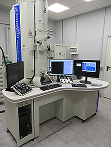
Laboratory for Nanoscopies and Spectroscopies-LANE
 |
Scientific responsible Dra. Cristina T. Rojas Ruiz · Dra. Asunción Fernández Camacho · Dr. Miguel Anaya Martín Technical staff Dra. Luisa María Valencia Liñan · Lda. Dª Olga Montes Amorín contact ✉ tcrojas@icmse.csic.es · olga@ciccartuja.es · asuncion@icmse.csic.es ☎ 954 48 96 25 |
The LANE laboratory has a TEM field emission microscope Tecnai F30, equipped with STEM mode, EDX and HAADF detectors, and an energy filter analyzer (GIF). In addition sample preparation equipment is available including plasma cleaning, surface polishing and thin films fabrication. The techniques available include: bright and dark field TEM; high resolution TEM; electron diffraction; STEM-HAADF analysis; EDX and STEM-EDX as well as EELS and STEM-EELS analysis (including point and line measurements and compositional maps); EFTEM images; spectrum-image and electron tomography analysis. More details about the LANE laboratory can be found on the website http://www.lane.icmse.csic.es/ (under construction).These facilities offer microstructural and chemical characterization especially devoted to the areas of materials science and physical chemistry of the solid state. It is relevant the high resolution analytical microscopy to characterize materials in the nano-scale..
Available Equipment
- Microscopio Tecnai G2 F30 S-TWIN 300KV con pistola de emisión de campo. Resolución estructural de 0,2 nm entre puntos, portamuestras de uno y dos ángulos. Adjuntos al equipo hay un detector de derivación de silicio EDX X-Max 80T Detector (Oxford Instruments) y un filtro de energía Gatan (GIF Quantum SE)
- Additional equipment in the “electron microscopy samples preparation laboratory”
|
Services
|
External | |
|---|---|---|
| OPI / AGE / Universidades | Others | |
| Structural Characterization: Tranmision Electron Microscopy (TEM) |
159,96 (€/hour)
|
183,95 (€/hour)
|
|
TEM: EDS (Energy Dispersive X-Rays Spectroscopy)
|
159,96 (€/hour)
|
183,95 (€/hour)
|
|
TEM: EELS (Electron Energy Loss Spectroscopy)
|
159,96 (€/hour)
|
183,95 (€/hour)
|
|
TEM: Spectrum imaging
|
159,96 (€/hour) |
183,95 (€/hour)
|
| TEM: High Resolution TEM | 159,96 (€/hour) | 183,95 (€/hour) |
| TEM: STEM/ADF-HAADF | 159,96 (€/hour) | 183,95 (€/hour) |
Electron Microscopy Samples Preparation Laboratory
 |
Scientific responsible Dra. Cristina T. Rojas Ruiz · Dra. Asunción Fernández Camacho Technical staff Dra. Luisa María Valencia Liñán · Dª María Inmaculada Rosa Cejudo · Lda. Dª Olga Montes Amorín contact ✉ tcrojas@icmse.csic.es |
The laboratory for TEM and SEM samples preparation has a gold coater, a carbon evaporator, a metallization system for Cr and carbon, a diamond wheel cutter, a grinder with disc-grinder device, an ultrasonic cutter, a concave polishing (dimple) and ion thinning (Fischione 1010).
|
Services
|
External | |
|---|---|---|
| OPI / AGE / Universidades | Others | |
|
Metalización (Sputtering)
|
41,20 (€/10 sample)
|
45,12 (€/10 sample)
|
|
Preparación de Láminas Delgadas y Probetas Delgado-Pulidas (Sección Transversal-Planar)
|
104,68 (€/sample)
|
114,65 (€/sample)
|
|
Preparación de Láminas Delgadas y Probetas Delgado-Pulidas (Adelgazamiento Iónico)
|
41,48 (€/sample)
|
51,36 (€/sample)
|
Preparation, characterization and processing of materials
Preparation, characterization and processing of materials
Preparation, characterization and processing of materials
| Scientific responsible Dr. Alfonso Caballero Martínez contact ✉ alfonso.caballero@csic.es ☎ 954 48 95 38 |
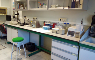
This Service can supply all types of solid samples with catalytic activity in various processes of industrial, energy and environmental interest.
Samples are supplied at any stage of preparation, with or without pretreatment or even ready to use. It can include their characterization by various physical and chemical techniques.
|
Services
|
External | |
|---|---|---|
| OPI / AGE / Universidades | Others | |
|
Evaluation of catalytic performance
|
91,12 (€/hour)
|
99,80 (€/hour)
|
|
Sample Preparation
|
91,12 (€/hour)
|
99,80 (€/hour)
|
| Thermal and chemical treatments | 91,12 (€/hour) | 99,80 (€/hour) |
Mechanized Workshop
Mechanized Workshop
Mechanized Workshop
 |
Scientific responsible Dr. Juan Pedro Espinós Manzorro Technical staff D. Juan Carlos Martín Sánchez · D. Manuel Perea Domínguez contact ✉ jcarlos.martin@csic.es ☎ 954 48 96 17 · int.: 446 102 |
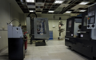
This is a fundamental horizontal service for the ICMS and external units attached to it. Since it allows the scientific material and equipment to be improved, modified and adapted to the needs of each researcher and/or ongoing research; arriving at its design and manufacturing based on a specific need, in addition to the technical advice of all the elements described above.
It also offers the possibility of carrying out small repairs and part of the general maintenance of the scientific and laboratory equipment of the ICMS.
Features and Instrumentation available.
The service has machine tools, manual and electric tools for the shaping of very diverse materials. According to the benefits offered, they are classified as follows:
CAD design of 2D or 3D parts.
In the technical office section for CAD design, there is Autodesk Fusion software.
Manufacture of parts using chip removal procedure.
For machining processes by chip removal, the following machine tools are available:
- CNC vertical milling machine, HAAS TM 1P.
- Conventional turret milling machine, Fortex FTX-4-FC VARIO.
- Vertical drill, ERLO TSAR32.
- Conventional parallel lathe, PINACHO SC200.
- Semi-automatic parallel lathe, PINACHO SMART TURN180.
- Band saw, Optimum S210G.
Rapid prototyping of parts by plastic deposit.
For rapid prototyping of parts made of various technical plastics, the following equipment is available:
- CoreXY FDM 3D Printer
High-performance 2D waterjet cutting.
For 2D CNC cutting by high-performance abrasive waterjet, the machine tool is
available:
- OMAX ProtoMAX abrasive waterjet cutting table.
Welding processes of metallic materials.
Fusion welding processes are used to join various metallic materials:
- Autogenous welding, strong and soft, with different contributions.
- MMA welding, electric arc.
- TIG welding on steels.
|
Services
|
External | |
|---|---|---|
| OPI / AGE / Universidades | Others | |
| Highñperformance 2D waterjet cutting (2D CN cutting) | 37,88 (€/minutes) | 46,89 (€/minutes) |
| CAD design of 2D or 3D parts | 32,01 (€/hour) | 39,63 (€/hour) |
| Manufacturing of parts by removal procedure (Machining with Lathe or Milling Machine) | 68,58 (€/hour) | 84,91 (€/hour) |
| Welding processes of metallic materials (Fusion Welding) | 110,52 (€/hour) | 136,83 (€/hour) |
| Rapid Prototyping of parts by plastic deposition (Manufacture of Plastic Parts 3D Printing) | 47,76 (€/hour) | 59,13 (€/hour) |
| Sandblasting | 61,19 (€/hour) | 75,76 (€/hour) |
icms